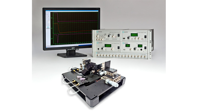
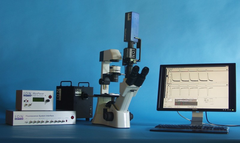
1500A:细小组织测量系统
一站式系统:测量完整细小肌肉或生理组织的各种力学性能
这是一个高度集成和一站式系统,其设计理念是提供一个简要的方法让研究者去控制和测量细小组织和肌肉的力学性能。
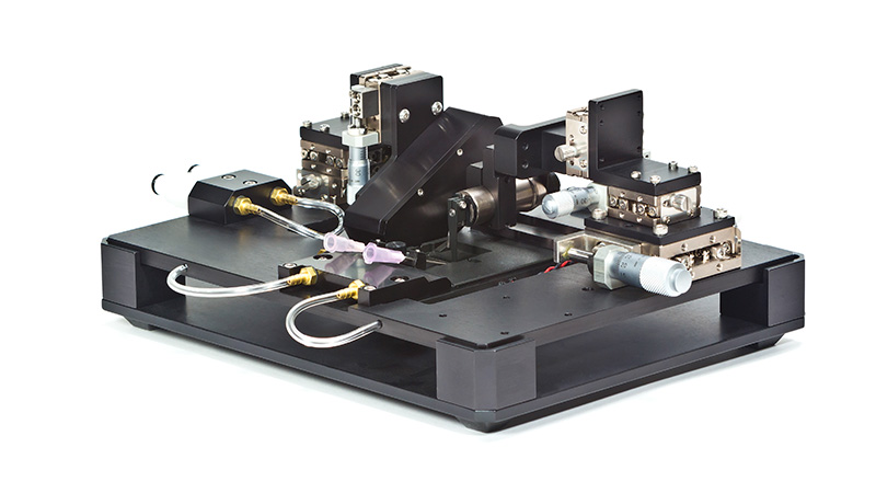
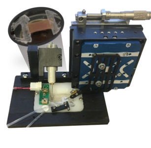
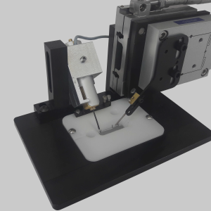
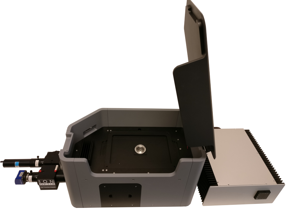
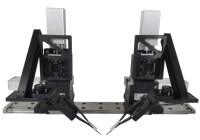
1500A,1510A和1530A细小组织测试系统让研究者能够在高可信度和可再现的基础下准确地测量细小组织的各种力学关系和参数,例如:长度-张力,力-收缩速率,功环和硬度。给予无限制的配置调整空间,三种不同的组装(1500A,1510A,1530A)具多功能性,能够精准地测量一些细小组织的力度和长度。每个组合的实验平台装置内置配有微米级XYZ位置调节器去调整我们400A系列的力传感器,322C高速长度操控仪和300C双模传感器的位置。
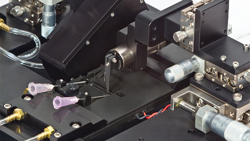
肌肉或组织透过浴槽边上独特的狭缝与力传感器的悬臂进行连结,加上使用适当的上盖置於浴槽顶部,整体设计便成为应用於人工肌肉结构物或耗氧实验的理想装置—耗氧实验需要封闭的环境。在系统运作下,样本的力度或长度都能够被测量或控制,从简单的张力到其他比较复杂的力学性能都可以测量出来。
信号采集和数据管理可以通过软件和标准化方案之间的配合去完成,把复杂的实验转化成简单和直接的测量。此外,系统亦提供一个金属底座让测试平台装置可安放在其上,方便存放,同时也令平台装置在底座和(立体或倒置)显微镜之间的移动更加安全和方便。
● 完整的测试系统且配有针对细小组织的浴槽平台装置
● 可控温的浴槽 (尺寸由400µL至1900µL)
● 能够测量和控制力度和长度
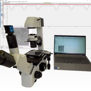
● 与标准显微镜和倒置显微镜兼容使用
● 可配置我们的肌小节间隔长度检测软件(HVSL/ VSL)
● 分辨率达至0.01µN
● 测量的力值峰度: 0.5mN至1000mN
新颖的浴槽设计
槽配备了溶液灌流装置,内置刺激电极和玻璃底部,能在保存组织存活同时对其进行成像观察。浴槽为横向设计和顶部敞开形,配合适当的上盖可形成封闭系统,以应用於耗氧或缺氧实验。
附加荧光和肌小节长度测量
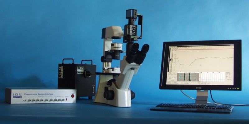
1500A兼容於市场上大部份的倒置显微镜,同时允可附加元件以测量肌小节长度和生物荧光。将荧光测量装置,肌小节间隔长度检测装置搭配在您的1500A上,就能具备实时和同时进行力度,肌节长度,钙浓度三方面的测量。
1500A系列是最灵活的系统,可以使用何种类型的样品。该系统有一个小型水平浴,适用于任何小型骨骼肌,心脏和平滑肌条,以及完整的单纤维和小束。还使用该系统进行了人工生长的肌肉构造的机械测量。使用该系统进行的实验类型类似于1200A和1300A(仅体外)系列,然而,1500A提供了额外的特征和补充实验,例如组织的生物荧光以及测量氧气消耗的潜力。在实验期间。
常见样品:
骨骼肌:趾长伸肌(EDL)、比目鱼肌、跖肌、横膈膜、蚓状肌、工程结构(iPSC 衍生)
心肌:单束或小束去膜纤维(通常是小梁或乳头肌束纤维)、工程结构(iPSC 衍生;sheets, bundles, rings)
平滑肌:单束或小束去膜纤维(通常是膀胱,结肠,阴道或维管束或纤维)、任何肌肉都可以测试
人造肌肉和结缔组织:软骨、上皮组织、肌腱
生物材料和聚合物:介质弹性体致动器(EAP)、电活性聚合物、凝胶和支架、电化学致动器、电热致动器
常见实验:
抽搐:设计用于引起单个或少量肌肉纤维收缩的单脉冲。

强直收缩:快速连续多次电脉冲,导致时间总和和完全肌肉收缩。

疲劳:经常重复次最大的强直性收缩,以引起肌肉疲劳。
力频率:改变刺激频率的速率,以评估引起最大强直作用力的最佳频率。
步态分析/建模练习(等渗,同心):控制肌肉力量输出(等渗)以评估肌肉的缩短速度。
刚度:被动正弦延长和肌肉缩短,以评估组织的固有刚度。
应力应变:增量延长组织以计算材料的杨氏模量。
力-速度(缩短速度):钙中纤维的最大活化,然后是一系列力钳,达到最大力的百分比,从而可以测量缩短的速度。
刚度:被动正弦延长和肌肉缩短,以评估组织的固有刚度。
主动力量长度(Frank Starling):以恒定或变化的频率起搏心脏组织,同时逐渐延长肌肉以增加预负荷,以评估兴奋收缩机制。
Norden, Diana M. et al. “Tumor growth increases neuroinflammation, fatigue and depressive-like behavior prior to alterations in muscle function.” Brain, Behavior, and Immunity (2015) DOI: 10.1016/j.bbi.2014.07.013
Tangney, Jared R. et al. “Timing and magnitude of systolic stretch affect myofilament activation and mechanical work.”” American Journal of Physiology-Heart and Circulatory Physiology (2014) DOI: 10.1152/ajpheart.00233.2014
Powers et al. “Subcellular Remodeling in Filamin C Deficient Mouse Hearts Impairs Myocyte Tension Development during Progression of Dilated Cardiomyopathy” International Journal of Molecular Sciences (2022) DOI: 10.3390/ijms23020871
Schick et al. “Reduced preload increases Mechanical Control (strain-rate dependence) of Relaxation by modifying myosin kinetics” Archives of Biochemistry and Biophysics (2021) DOI: 10.1016/j.abb.2021.108909
Schick et al. “Reduced preload increases Mechanical Control (strain-rate dependence) of Relaxation by modifying myosin kinetics” Archives of Biochemistry and Biophysics (2021) DOI: 10.1016/j.abb.2021.108909
Zuo, Li, Leonardo Nogueira, and Michael C. Hogan. “Reactive oxygen species formation during tetanic contractions in single isolated Xenopus myofibers.” Journal of Applied Physiology (2011) DOI: 10.1152/japplphysiol.00398.2011
Zhang et al. “Maturation of human embryonic stem cell-derived cardiomyocytes (hESC-CMs) in 3D collagen matrix: Effects of niche cell supplementation and mechanical stimulation” Acta Biomaterialia (2017) DOI: 10.1016/j.actbio.2016.11.058
Zuo, Li et al. “Low Po2 conditions induce reactive oxygen species formation during contractions in single skeletal muscle fibers.” American Journal of Physiology-Regulatory, Integrative and Comparative Physiology (2013) DOI: 10.1152/ajpregu.00563.2012
Gharanei, Mayel “Investigation into the cardiotoxic effects of doxorubicin on contractile function and the protection afforded by cyclosporin A using the work-loop assay.” Toxicology in Vitro (2014) DOI: 10.1016/j.tiv.2014.01.011
Laurila et al. “Inhibition of sphingolipid de novo synthesis counteracts muscular dystrophy” Science Advances (2022) DOI: 10.1126/sciadv.abh4423
Blitz et al. “Infiltration of intramuscular adipose tissue impairs skeletal muscle contraction” The Journal of Physiology (2020) DOI: 10.1113/JP279595
Bailey et al. “Incubation with sodium nitrite attenuates fatigue development in intact single mouse fibres at physiological” The Journal of Physiology (2019) DOI: 10.1113/JP278494
Ferrera et al. “Hypothermia Decreases O2 Cost for ex vivo Contraction in Mouse Skeletal Muscle” Medicine and Science in Sports and Exercise (2018) DOI: 10.1249/MSS.0000000000001673
Tangney, Jared R. “Effects of alterations in sarcomere structure and prestretch timing on cardiac muscle mechanics.” MSc Thesis. University of California San Diego (2012) DOI: N/A
Rhim, Caroline et al. “Effect of MicroRNA Modulation on Bioartificial Muscle Function.” Tissue Engineering (2010) DOI: 10.1089/ten.TEA.2009.0601
Brynnel et al. “Downsizing the molecular spring of the giant protein titin reveals that skeletal muscle titin determines passive stiffness and drives longitudinal hypertrophy” eLife (2018) DOI: 10.7554/eLife.40532
Nogueira et al. “Cigarette smoke directly impairs skeletal muscle function through capillary regression and altered myofibre calcium kinetics in mice” The Journal of Physiology (2018) DOI: 10.1113/JP275888
Hall et al. “Cellular rescue in a zebrafish model of congenital muscular dystrophy type 1A” NPJ Regenerative Medicine (2019) DOI: 10.1038/s41536-019-0084-5
de Lange, Willem J. et al. “Ablation of cardiac myosin-binding protein-C accelerates contractile kinetics in engineered cardiac tissue.” Journal of General Physiology (2013) DOI: 10.1085/jgp.201210837


